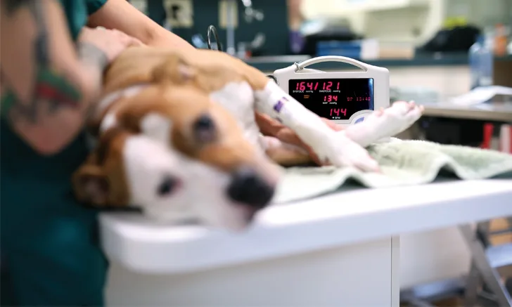
Blood Pressure Measurement
Oriana D. Scislowicz, LVT, aPHR, CVCA-Cardiac Care for Pets, Glen Allen, Virginia

Blood pressure measurement is an essential veterinary nursing skill that is used as a diagnostic tool in a multitude of veterinary settings, from emergency and critical care environments where patients require constant monitoring to general practice settings where patients receive baseline health assessments.
In an anesthetic setting, constant evaluation of a patient’s blood pressure helps avoid serious complications from sustained hypertension such as eye, ear, brain, and kidney damage. When blood pressure is markedly elevated during an anesthetic event, the patient may be at risk for development of end-organ damage or vascular incidents.2 Hypotensive patients under anesthesia may experience damage to the kidneys and other vital organs.2
In general practice, many diseases and drugs can result in blood pressure changes, so measurement is an important regular diagnostic tool during examination. Diabetes, hyperadrenocorticism, hyperthyroidism, and renal, hepatic, and cardiac insufficiencies can all cause hypertension.3
Some drugs (eg, the methylxanthine family [minophylline, theophylline]) can also result in hypertension. Moreover, patients presenting with certain clinical signs (eg, dilated pupils, hyphema, weight loss, hematuria, heart murmurs) may be experiencing occult hypertension if blood pressure measurement is not regularly assessed.3
Blood Pressure Physiology
Blood pressure drives perfusion, which is the delivery of blood and oxygen to organs (eg, brain, heart, lungs, kidneys), as well as tissue beds within the body. Blood pressure values come from the left ventricular contraction that pushes blood into the aorta, which creates the systolic arterial pressure (SAP). The left ventricle then empties and relaxes, and aortic pressure falls as the left ventricle fills again, which results in the diastolic arterial pressure (DAP).
The equation for determining the mean arterial pressure (MAP) uses both the diastolic and systolic arterial pressure readings1:
MAP = DAP+1/3 (SAP-DAP)
Values are measured in millimeters of mercury (ie, mmHg).
Understanding the roles that cardiac output and systemic vascular resistance play in the MAP values is important because many drugs used for anesthesia affect these in some fashion. (See Resources.)
Measurement Techniques
Arterial blood pressure can be monitored directly or indirectly. Direct arterial blood pressure monitoring, considered the gold standard, provides continuous monitoring of systolic, diastolic, and MAP via an arterial catheter.1 Indirect arterial blood pressure monitors, which are more commonly used in daily practice, include Doppler and oscillometric methods that measure arterial blood flow in a peripheral artery. (See Standard Operating Procedures for Blood Pressure Measurement.)
Direct Monitoring
In direct blood pressure monitoring, a catheter is placed into an artery and attached to a transducer, which is placed at the level of the right atrium. Continuous systolic, mean, and diastolic readings are obtained via an oscilloscope that is connected to the transducer. Catheters are most commonly placed in the palmar arterial arches of the thoracic and pelvic limbs, although other potential sites include the femoral and auricular arteries. Sterile technique should be used during placement to reduce the risk of complications (eg, hematoma, air embolism, infection, thrombosis). The catheter should be clearly marked so it is not mistaken for an intravenous catheter, and only heparinized saline should be injected into it. The catheter should be flushed regularly with heparinized saline.4
Indirect Monitoring
In Doppler monitoring, one indirect blood pressure measurement method, arterial wall motion is detected by the ultrasound waves created from the transducer. The Doppler effect occurs when a frequency change from a moving object results in the reflected sound beam reaching an audible range from a previous ultrasonic range.4 In the case of Doppler blood pressure monitoring, the moving object is turbulent blood flow within the vibrating arterial walls. In human medicine, the nurse is able to listen for these vibrations with a stethoscope, whereas in veterinary patients, these sounds (ie, Korotkoff sounds) are inaudible. This makes it necessary to use the Doppler transducer to detect blood flow instead of relying on listening via a stethoscope.5
Similar to direct blood pressure monitoring sites, the palmar arterial arches of the fore- and hindlimbs are ideal locations.4 For cats that do not cooperate with fore- or hindlimb measurement, the veterinary nurse may place the transducer against the coccygeal artery located on the ventral aspect of the tail, near the tail base. When selecting a cuff, choose one with a short axis that is approximately 40% of the circumference of the limb or tail being used to obtain the reading. A cuff that is too large may cause falsely low readings, whereas cuffs that are too small may lead to artificially high readings. Limb placement is also very important, as limbs should be as close to heart level as possible. When cuffs are placed on limbs well above or below the heart, false low or false high readings, respectively, may occur.4
Standard Operating Procedures for Blood Pressure Monitoring
Direct Blood Pressure Monitoring
Supplies needed:
Heparinized saline
IV catheters
Luer lock 3-way stopcock
Small-diameter pressure tubing
Pressure transducer
Direct blood pressure monitor and cable
Remove the catheter end-cap using heparinized saline to flush the catheter. Slide the catheter up the stylet to flush the inner catheter.
Flush the 3-way stopcock with heparinized saline.
Fill the transducer with heparinized saline, making sure that no air bubbles are present.
Flush through the perpendicular arm of the 3-way stopcock and attach pressure tubing to the stopcock.
Transfer the heparinized saline syringe to the perpendicular port. Rotate the valve and flush with heparinized saline to direct the flow into the tubing.
Palpate the arterial site (ie, most commonly, the dorsal pedal artery), sterilely prepare the site by clipping the area and scrubbing as you would for IV catheter placement, and aseptically place the catheter in the artery.
Ensure the pressure transducer is secured to the monitor cable. Attach the 3-way stopcock and tubing and secure into place. Attach the opposite end of the tubing to the pressure transducer.
Select ART on the direct blood pressure monitor.
Rotate the valve, then zero the transducer and maintain it at heart base level to expose the transducer chamber to normal atmospheric pressure.
Attach a heparinized saline syringe to the open 3-way valve port at the transducer. The valve should then be rotated to close flow to the transducer and the tubing and the catheter should be flushed. Rotate the 3-way valve to allow connection between the arterial catheter and transducer chamber.
When a well-defined wave form is observed on the monitor, zero the transducer and maintain it at heart base level.
Flush the fluid line intermittently when the pulse wave diminishes to prevent clots from developing.
Indirect Blood Pressure Monitoring: Doppler
Supplies needed:
Doppler blood pressure device
White porous tape
Ultrasound transmission gel
Clippers
Sphygmomanometer
Blood pressure cuffs
Headphones (optional)
Measure the short axis of the cuff, which should be approximately 40% of the patient’s appendage circumference. Apply the cuff to the selected limb or tail.
Apply a generous amount of ultrasound transmission gel to the transducer, which should then be applied to the caudal metacarpal or metatarsal area, or over the coccygeal artery at the base of the tail. If needed, shave a small amount of fur from the site. Turn the Doppler machine on to check for a signal.
Wrap 1-inch white porous tape around the transducer and the limb or tail with moderately firm pressure for sustained monitoring under anesthesia.
Attach the sphygmomanometer to the hose on the cuff. Pump the pressure up until the signal is lost.
Release pressure gradually, listening for the return of the signal that corresponds with the systolic blood pressure reading.
Indirect Blood Pressure Monitoring: Oscillometric
Supplies needed:
Oscillometric monitor & adapter tubing
Blood pressure cuffs
Measure the short axis of the cuff, which should be approximately 40% of the patient’s appendage circumference. Apply the cuff to the selected limb or tail.
Attach the cuff tubing to the monitor’s adapter tubing.
Turn the oscillometric monitor on, press “start,” allow the monitor to begin inflating the cuff, and obtain a reading. The monitor will provide a diastolic, systolic, and mean arterial blood pressure reading.
After a cuff is selected, the area can be shaved or the fur parted, and a generous amount of ultrasonic gel applied to the area or the transducer. The transducer is placed against an artery with the concave side facing the skin surface. The cuff is placed above the transducer, and a sphygmomanometer is used to inflate the cuff. For long-term measurement (eg, during anesthetic monitoring), the transducer can be secured in place with porous tape. After the cuff is inflated, allow it to slowly deflate until blood flow returns. The systolic measurement will be heard as the first audible sound; once the sound is heard, the blood pressure measurement will be read from the sphygmomanometer.4
The same measurement and placement of blood pressure cuffs are used in oscillometric methods, but with a device that measures oscillations within the cuff bladder, without the use of a transducer or sphygmomanometer. The cuff is slowly inflated and deflated automatically by the machine. The device then provides the systolic, mean, and diastolic readings, along with a heart rate. A 2004 study comparing Doppler and oscillometric blood pressure monitoring methods in feline patients found that the Doppler method was well correlated with direct blood pressure measurements, while the oscillometric device was poorly correlated, demonstrating the increased reliability of the Doppler method compared with oscillometry.6
Conclusion
Blood pressure measurement is a relatively simple diagnostic procedure that can provide a wealth of information to help assess disease processes and assist in successful anesthetic monitoring of veterinary patients. When used as a routine diagnostic tool during physical examinations, hypertension and underlying disease processes can be caught earlier or at onset. In the anesthetic realm, hypotension and hypertension are also easier to manage when caught early and treated appropriately with fluid and drug therapy.
Letter to the Editor
This article originally appeared in the October 2018 issue of Veterinary Team Brief.