Urethral obstruction is common in male cats and can be idiopathic or caused by urethral mucous plugs, urolithiasis, strictures, or neoplasia.1,2 Recommended treatment is typically urethral catheterization.1,2 Depending on the duration of obstruction, stabilization may be needed prior to administration of sedation or anesthesia to facilitate placement of a urinary catheter.
Azotemia, electrolyte abnormalities (eg, hyperkalemia), acidemia, and cardiovascular events (eg, arrhythmias) are the most common comorbidities at presentation in obstructed cats. In a study of 168 cats, 57% had azotemia, 46% had hyperkalemia, 73% had acidemia, and 33.5% had arrhythmias; arrhythmias were primarily bradycardia (88.5%) and ventricular premature complexes (11.4%; see Step 4).3 Absence of P waves with normal QRS complexes (ie, atrial standstill) is also common in blocked cats with hyperkalemia.4 Less commonly, cats can be presented with hypovolemia and hypotension or significant clinical dehydration (53% of cats in one study).4
The goal of stabilization is to identify and address expected comorbidities to optimize the patient for safe sedation or anesthesia prior to urinary catheter placement. Before sedation or anesthesia, cats should be normovolemic with normal blood pressure, have normal sinus rhythm on ECG, and receive treatment for hyperkalemia if potassium is >7 mEq/L (7 mmol/L). Blocked cats will not be completely stable until a urinary catheter is placed. Unblocking should typically be attempted prior to referral for further care and hospitalization.
When a patient is critically ill, it can be especially tough to talk about money, but finances can be the deciding factor in patient care. Set these conversations up for success.
Provide a safe place for clients in a private room (not the waiting area), and have a seat together.
Acknowledge the client's financial strain (if applicable) and offer partnership in seeking solutions.
Ask permission before offering suggestions.
Present small amounts of information and pause to let the client absorb and formulate questions.
Include the rationale and benefit to the patient for the diagnostics and treatments you anticipate.
Step-by-Step: Stabilization of Cats With Urethral Obstruction Prior to Referral
What You Will Need
IV catheter
Crystalloid fluid (typically a buffered isotonic crystalloid)2
Monitoring units
ECG
Blood pressure unit (ultrasonic Doppler or oscillometric unit)
Blood gas analyzer or other machine capable of measuring electrolytes and, ideally, either blood pH or total carbon dioxide to estimate metabolic acidosis
Pharmacologic interventions
Calcium gluconate
50% dextrose
Regular (short-acting) insulin
Bicarbonate
Opioid pain medication (eg, buprenorphine, methadone) ± sedative (eg, acepromazine, midazolam, dexmedetomidine, ketamine)
Urinary catheter
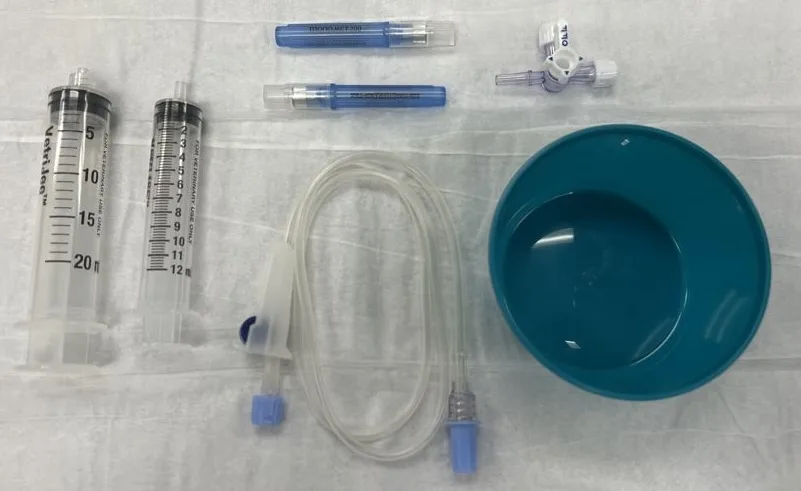
Decompressive cystocentesis supplies
Hypodermic needle (22 gauge, 1 or 1.5 inch)
Stopcock
IV extension set
12- or 20-mL syringe
Collection container for urine
Forced-air warmer, circulating hot water blanket, and/or blankets


Step 1: Assess the Patient
Perform a physical examination to determine whether the cat is in shock and identify any cardiovascular abnormalities (eg, murmurs, arrhythmias) before giving fluid therapy.


Author Insight
Palpating pulse quality, performing blood pressure measurement, and assessing heart rate and rhythm, mucous membrane color, rectal temperature, and general mentation can help diagnose shock. Most blocked cats are not in shock. The author does not routinely perform blood pressure measurement unless a patient is obtunded, laterally recumbent, and bradycardic (heart rate, <160 bpm).
Step 2: Place an IV Catheter, Warm the Patient, & Administer Analgesics
Place an IV catheter to facilitate drug administration (eg, analgesia, anesthesia), fluid therapy, and blood sampling. Warm hypothermic cats with a forced-air warmer, circulating hot water blanket, and/or regular blankets. Administer pain medication as indicated, but do not administer NSAIDs, as cats with urethral obstruction are often dehydrated (and/or hypovolemic) and azotemic.

Author Insight
Methadone (0.1-0.2 mg/kg IV) or buprenorphine (0.01-0.03 mg/kg IV) is commonly administered for analgesia. With buprenorphine, onset of analgesic effects can take ≥20 to 30 minutes.
In the author’s experience, most cats allow IV catheter placement prior to drug administration, but painful, anxious, or fractious cats may require IM administration of opioid pain medications and sedatives to facilitate catheterization. Ideally, patients without electrolyte and ECG results should only be given opioids and benzodiazepines, but ensuring patient compliance is key because IV catheterization is important for definitive treatment (ie, placement of a urinary catheter). The author therefore administers methadone (0.1 mg/kg IM) with or without midazolam (0.2 mg/kg IM) in mildly fractious cats and methadone (0.1 mg/kg IM), dexmedetomidine (5-7 micrograms/kg IM), and ketamine (2 mg/kg IM) in moderately to severely fractious cats in an attempt to provide sufficient sedation for management with a single injection. Dexmedetomidine should be administered with caution in very sick or bradycardic cats and reserved for fractious patients.
Step 3: Acquire a Blood Sample
Ideally, perform a full serum chemistry profile and CBC. If these analyses are not practical because of the time and volume of blood required, perform a packed-cell volume/total protein measurement and a limited panel that contains BUN, creatinine, and electrolytes (primarily potassium but also sodium and chloride). Measure pH directly or estimate by the total carbon dioxide or bicarbonate if available.
Author Insight
In the author’s experience, collecting a blood sample from an unflushed IV catheter at the time of catheter placement is easiest, but blood can be collected from any vessel.

Potassium >7 mEq/L (7 mmol/L) typically requires treatment (see Step 5), especially in patients with bradycardia or other arrhythmias or severe illness.4,5 Azotemia alone does not require direct treatment, as the prerenal component of azotemia improves with fluid therapy, and postrenal effects are corrected by urethral catheterization.6 Metabolic acidosis in obstructed cats is primarily due to azotemia, hyperphosphatemia (if present), and lactic acidosis and does not require direct treatment.
Step 4: Perform ECG
Perform ECG to determine whether an arrhythmia is present.

If an arrhythmia attributed to hyperkalemia (ie, bradycardia [Figure 1], atrial standstill [Figures 2 and 3]) or a ventricular arrhythmia is present with potassium >7 mEq/L (7 mmol/L), administer treatment for hyperkalemia (see Step 5). Reassess the arrhythmia following treatment to determine whether it has resolved.
In less common situations in which a ventricular arrhythmia (intermittent ventricular premature complexes or sustained ventricular beats) is noted with potassium <7 mEq/L (7 mmol/L) or after treatment of hyperkalemia, administer fluids (typically, 10-20 mL/kg bolus) to improve tissue oxygen delivery and hypotension (if present) in addition to flow-by oxygen therapy. If the arrhythmia and a heart rate >180 to 200 bpm (ie, ventricular tachycardia) persist following fluid and oxygen therapy, administer a bolus of lidocaine (0.2-0.5 mg/kg IV).

Sinus bradycardia (heart rate, 126 bpm) with P waves and deep, negatively deflected T waves

Atrial standstill with one ventricular premature complex (arrow), negatively deflected T waves, and absent P waves

Atrial standstill with one ventricular premature complex (arrow), positively deflected T waves that are as tall as QRS complexes, and absent P waves
Author Insight
ECG is ideally performed prior to administration of sedatives or anesthetic drugs; however, sedation or anesthesia may be needed to obtain ECG in fractious patients.
The author does not always perform ECG in cats that are clinically bright and alert and have a heart rate >180 bpm at presentation.
The author always administers a fluid bolus (5-10 mL/kg IV over 15-30 minutes) to provide fluids prior to and during sedation/anesthesia and replace mild dehydration.
Step 5: Treat Hyperkalemia
If hyperkalemia is present, administer IV fluids (see Step 6) and select a cardioprotective medication that reduces blood potassium levels or improves cardiomyocyte function (Table 1).
Table 1: Drugs for Treatment of Hyperkalemia
Author Insight
The author prefers calcium gluconate, as administration of dextrose and regular (short-acting) insulin can induce acute or delayed (hours later) hypoglycemia, and the duration of effect of calcium gluconate is typically sufficient to unblock the cat (ie, provide definitive treatment for hyperkalemia).
Step 6: Administer Fluid Therapy
Administer buffered isotonic crystalloid fluids as appropriate to help increase the glomerular filtration rate and promote excretion of potassium, BUN, and creatinine through the kidneys (Table 2).
Table 2: Suggested Fluid Therapy for Common Scenarios in Blocked Cats
Author Insight
A murmur or gallop rhythm may indicate underlying heart disease. In these cases, lower maintenance fluid rates (40 mL/kg/day CRI) should be used, and dehydration deficits should be replaced over ≥24 hours in stable patients. When bolusing fluids in hypovolemic cats with suspected heart disease, smaller boluses (5-10 mL/kg) or administration over longer periods of time (20-30 minutes) should be considered.
When making a referral recommendation, include the client in the decision, and consider whether the client is willing to seek care at another facility and whether additional treatment will be cost-prohibitive.
Must Reads From Our Newsletter:
Step 7: Prepare Patient for Referral
Attempt to place a urethral catheter. If unsuccessful, determine whether the patient can be referred without catheterization and whether decompressive cystocentesis is needed.
Author Insight
Urethral Catheterization Prior to Referral (Preferred)
Cats should ideally be unblocked via placement of a urethral catheter prior to referral for 24-hour hospitalization and monitoring, especially cats that are hyperkalemic at presentation. Patients should be transferred with the urinary catheter and collection bag.
Review this guide for urethral deobstruction via catheterization in cats.
Referral Without Urethral Catheterization
If the referral clinic is ≤2 hours away and the cat is not hypotensive, bradycardic, or hyperkalemic, the patient may be referred without catheterization if catheterization is unsuccessful, thus avoiding potential complications of decompressive cystocentesis (eg, uroabdomen, trauma to the bladder wall). If pain medications were not previously administered as part of IV or urethral catheterization attempts, an opioid pain medication can be administered (see Step 2).
Decompressive Cystocentesis Prior to Referral
If the referral clinic is >2 hours away or the cat was hypotensive, bradycardic, and/or hyperkalemic at presentation, decompressive cystocentesis should be performed to maintain patient stability during transit (see Step-by-Step: Decompressive Cystocentesis). Pet owners should be informed of risk for bladder trauma and/or uroabdomen with decompressive cystocentesis. Uroabdomen is typically identified by the referral clinic prior to urethral catheterization or after manipulating a damaged bladder during urethral catheterization.
Step-by-Step: Decompressive Cystocentesis
Step 1
Administer sedation if needed to prevent patient movement and minimize the likelihood of bladder trauma.
Step 2
Place a 22-gauge, 1- or 1.5-inch needle in the center of the urinary bladder at a 30- to 90-degree angle to the body using palpation (blind) or ultrasound guidance. If needed, use one hand to stabilize the urinary bladder while the needle is inserted.
Step 3
Attach the needle to an extension set, stopcock, and syringe.

Step 4
Drain as much urine as possible from the bladder.
Author Insight
A 1.5-inch needle helps ensure the needle remains in the urinary bladder as the bladder shrinks during emptying. Repeatedly puncturing the bladder should be avoided to reduce the likelihood of compromising the bladder wall, which can lead to uroabdomen.
Make transfers to the emergency room (ER) less stressful for both teams by following some of the following suggestions from ER clinicians.
Make the phone call yourself. It is easier to understand a patient's condition when speaking directly with the clinician.
Prepare the client with a cost estimate, prognosis, and estimated wait time, and document the conversation.
Communicate with the ER clinician about client conversations and expectations.
Share records electronically, and send a physical copy with the client.
Provide accurate times for vitals, dosages, and treatments.
Listen to The Podcast
Dr. Thomovsky highlights the importance of recognizing early signs of obstruction, performing prompt physical examinations, and managing associated electrolyte imbalances, including the emergency treatment of hyperkalemia.