Stella, a 6-year-old, 55-lb (25-kg) spayed American pit bull terrier crossbreed, was presented for intense pruritus (pruritus visual analog scale, 10/10) and erythematous nodules of 8-weeks’ duration. On presentation 8 weeks prior, nodules were thought to be hives. Treatment with oclacitinib (0.5 mg/kg PO every 24 hours) and cephalexin (22 mg/kg PO every 12 hours) for 4 weeks failed to improve clinical signs. Prednisone (0.5 mg/kg PO every 24 hours) was then administered for 2 weeks, but pruritus remained unchanged, and nodules continued to progress.
Physical Examination
On physical examination, Stella was quiet, alert, and responsive. BCS was 5/9, with no significant changes since the previous visit. No abnormalities were noted on thoracic auscultation, vital parameters were within normal limits, and lymph nodes were not enlarged.
Dermatologic Examination
Multiple multifocal, firm, erythematous nodules (diameter, 0.5-4.5 cm) were present over the entire body (Figures 1 and 2). Some lesions were eroded and ulcerated with crusting. An ulcerated nodule (0.4 cm × 0.2 cm) was present on the buccal surface of the left maxillary lip. New nodules that appeared following initial presentation were small (diameter, 0.1-0.5 cm), haired, and nonulcerated.
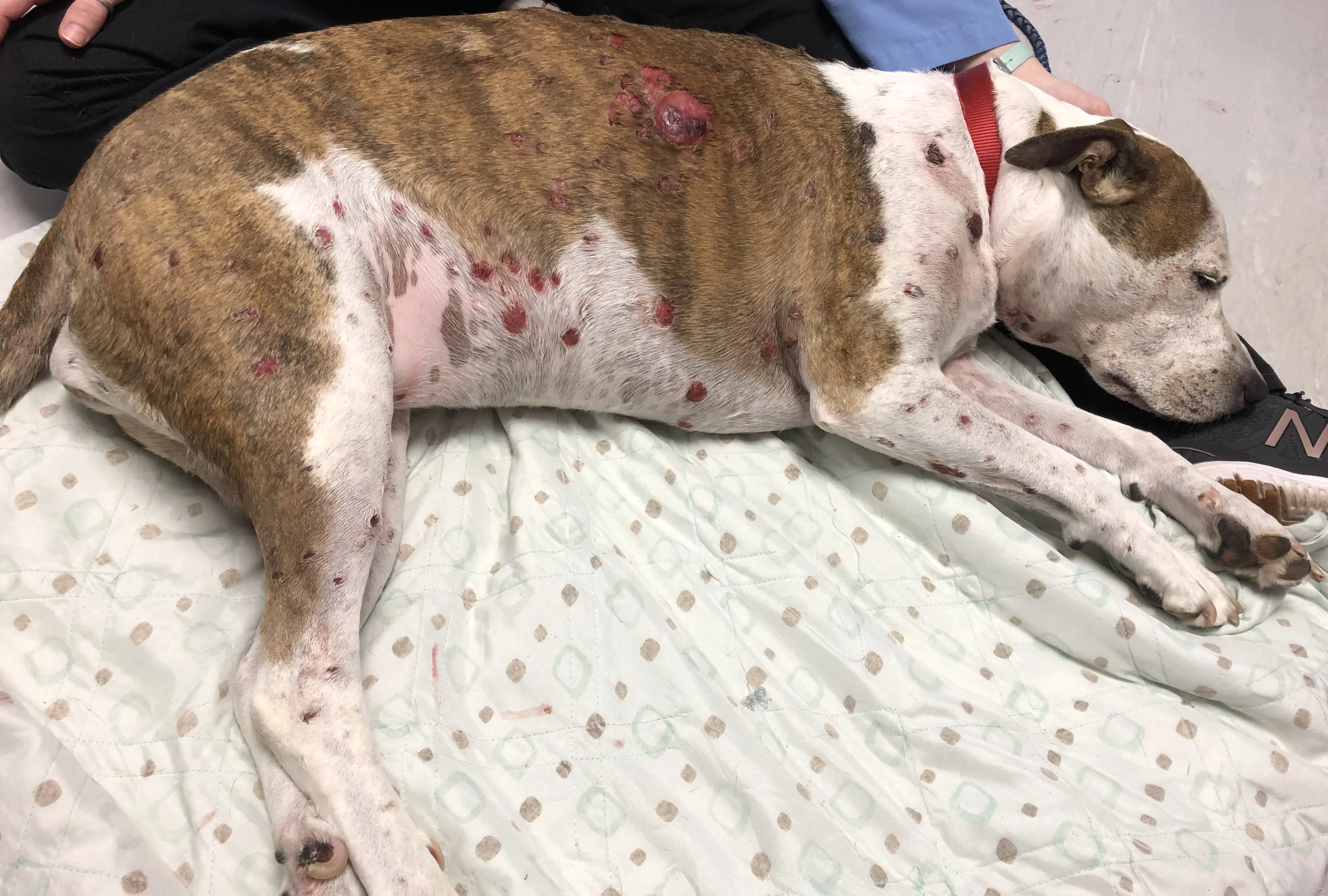
Multifocal cutaneous lesions

Erythematous, firm, cutaneous nodules
Diagnostics
Initial differential diagnoses included cutaneous neoplasia (epitheliotropic lymphoma, mastocytosis), histiocytic disease, infectious granulomatous disease, sterile or infectious panniculitis, deep pyoderma, and sterile pyogranulomatous dermatitis.
Impression smear cytology performed on multiple nodules showed RBCs and neutrophils. CBC and serum chemistry profile were unremarkable.
Stella was sedated with dexmedetomidine (3 micrograms/kg IV). Three sites were selected for biopsy, and 0.5 mL lidocaine (20 mg/mL) was administered at each site as a local anesthetic. Three 6-mm punch biopsies were performed on nodules at varying stages. Each biopsy site was closed with a single inverted cruciate suture using nonabsorbable suture material.
Histopathology showed round cells in the dermis, subcutis, follicular epithelia, apocrine glands, and epidermis (Figure 3) that occasionally formed small intraepidermal nests (ie, Pautrier's microabscesses, which are enlarged, atypical T-lymphocytes forming aggregates or found diffusely within the epidermis). Neoplastic round cells had distinct cell margins and central nuclei with finely stippled, normochromatic chromatin and 1 to 2 small nucleoli. Mitoses ranged from 3 to 16 per high-power field (40×), with 64 mitotic figures counted in 10 randomly selected high-power fields.

Histopathology showing infiltration of the epidermis by neoplastic lymphocytes. Image courtesy of Dr. Shannon Martinson
Diagnosis: Cutaneous Epitheliotropic T-Cell Lymphoma
Treatment & Long-Term Management
Surgery was not considered because multifocal nodules were present. Lomustine (CCNU; 60 mg/m2 PO every 3 weeks) was administered.1 Lomustine is a common treatment for cutaneous epitheliotropic T-cell lymphoma (CTCL) and is generally administered every 3 to 4 weeks as a chemotherapeutic medication. CBC and serum chemistry profile are recommended prior to each dose, as well as a CBC including platelet count 1 week after initial administration.2 Blood work monitoring is recommended because of potential serious adverse effects, including myelosuppression and hepatotoxicity.3,4 CBC at day 7 was unremarkable.
Treatment at a Glance
Multiple treatment options, including lomustine and surgical removal of single lesions, are available.
Blood work monitoring is important when administering chemotherapeutic agents because of potential for serious adverse effects, including myelosuppression and hepatotoxicity.
Secondary infections should be identified via cytology and treated appropriately.
Prognosis & Outcome
Skin lesions and pruritus were not responsive to treatment. The owners elected euthanasia 4 weeks after diagnosis.
Discussion
CTCL is an uncommon neoplasia in dogs (<1% of cutaneous tumors5), characterized by infiltration of neoplastic T lymphocytes with a tropism for the epidermis and adnexal structures.6,7 Etiology is unknown. Dogs with a history of atopic dermatitis can be 12 times more likely to develop CTCL,8 possibly due to chronic activation and proliferation of T lymphocytes that leads to clonal expansion.9
Clinical signs can vary, with 4 primary clinical presentations generally observed: pruritic erythema and scaling; mucocutaneous erythema, infiltration, depigmentation, and ulceration; solitary or multiple plaques or nodules; and infiltrative and ulcerative oral mucosal disease.3,6,10 Affected areas include haired skin (90%), lips (42%), the nasal planum (28%), and paws (27%).11 One study noted >50% of dogs had generalized scale (exfoliative dermatitis), <50% had plaques or nodules, and 30% had mucocutaneous lesions.12 Another study found diffuse erythema in 86.6% of cases, scaling in 60%, and focal hypopigmentation in 50%.6 Footpads may be hyperkeratotic, depigmented, or ulcerated.7,10 Systemic signs of illness (eg, lethargy, GI disease, anorexia) and lymphadenopathy may be present, and mucocutaneous involvement can occur in up to 50% of patients.6 The literature suggests there are no breed predispositions, but English cocker spaniels and boxers may be overrepresented.6,10 Average age at onset is 9 to 12 years.9,10 Anecdotally, most dogs with CTCL are intensely pruritic. One retrospective study, however, noted pruritus in only 40% of cases.6
Blood work is often unremarkable unless Sézary syndrome (neoplastic lymphocytes in the blood) is present.6,10 Metastasis is uncommon, with rare reports of disease in bone marrow, lymph nodes, and the spleen.13 Cytology of lesional skin may show round cells that may not be atypical and cannot be differentiated from clonal expansion.9 Definitive diagnosis includes skin biopsies and histopathology.
Inflammation is characterized by infiltration of neoplastic T lymphocytes with a tropism for the epidermal or mucosal epithelium, as well as adnexal structures, especially the follicular wall (Figure 4).7,12 Pautrier’s microabscesses (intraepithelial neoplastic lymphocytes diffusely in the epidermis or in aggregates) may be noted.7 Neoplastic lymphocytes that infiltrate apocrine sweat glands occur in 70% of cases and are considered diagnostic (Figure 5).6,7

Neoplastic lymphocytes infiltrating the follicular wall. Image courtesy of Dr. Shannon Martinson

Neoplastic lymphocytes infiltrating an apocrine sweat gland. Image courtesy of Dr. Shannon Martinson
Several treatment options have been documented and are available, including surgical removal of solitary lesions, lomustine, doxorubicin, L-asparaginase, dacarbazine, retinoids, radiotherapy, oclacitinib, and safflower oil.1,5,9,10,14-23 Response to treatment (ie, improvement in clinical signs and pruritus, complete remission or improvement) occurs in 78% to 83% of cases overall.6
Surgical removal is an option for solitary lesions, with 50% to 70% of dogs with single cutaneous or mucocutaneous lesions not developing further lesions for a median of 501 to 691 days.5 Staging should be performed prior to surgery to document any metastasis and to guide the final decision on whether to perform surgical removal. Systemic chemotherapy may also be needed, depending on margins obtained.15
Lomustine is often used and appears to be valuable in the treatment of CTCL. In a retrospective study, the response rate to lomustine was 78% (6 complete responses, 22 partial responses).18 Dogs received a median dose of 70 mg/m2 and a median number of 3 treatments. In a study of 46 dogs receiving lomustine, 15 dogs achieved complete response, 23 achieved partial response, 5 had stable disease, and 3 had progressive disease. Overall response rate was 83%, with a median duration of response of 94 days (range, 22-282 days).12 Adverse effects include increased liver enzymes (86%), GI signs (22%), and myelosuppression (29%).15,18 Blood work should be monitored during therapy and can increase the cost of treatment. Lomustine can be used in combination with other medications (eg, prednisone, other chemotherapeutic agents). In one study, 5 dogs received a single dose of L-asparaginase at the initiation of therapy with lomustine12; however, determining from studies whether combination therapies lead to better remission rates can be challenging.
Other reported treatments include radiotherapy for local treatment of neoplasia, L-asparaginase, doxorubicin, retinoids, and safflower oil, with response rates ranging from 42% to 75%.16,20-22 Occasional case reports document improvement in clinical signs with treatments like dacarbazine and oclacitinib.19,23 Therapies like retinoids can be expensive and challenging to obtain and thus are often not selected for treatment despite promising response rates. Further studies are needed to recommend some treatments (eg, oclacitinib) due to low numbers in published studies. Systemic prednisone does not appear to induce remission.1
CTCL is a progressive disease. Prognosis is difficult to establish and should be based on severity and treatment options. Mean survival time can be a few months to 2 years.6,9,10 Without treatment, survival time can be as low as 3 to 5 months. In one study, survival time was not significantly different between treated and untreated dogs.1 Cutaneous form of CTCL and multiple lesions are predictors of poor survival.1 Complete remission with treatment is associated with a longer survival time.5 Disease progression often results in euthanasia of affected patients.9
Take-Home Messages
Clinical presentation of CTCL is variable and called the great mimicker.
Histopathology of skin biopsies is the optimal diagnostic tool.
CTCL should be considered as a differential in atopic patients that stop responding to treatment.
Blood work is often unremarkable, and metastasis is rare.
Prognosis is poor, with many patients euthanized due to disease progression or lack of response to treatment.