Measuring Vertebral Left Atrial Size in Dogs
Erica Castaneda, DVM candidate, University of Florida
Amara H. Estrada, DVM, DACVIM (Cardiology), University of Florida

Vertebral left atrial size (VLAS) is a radiographic measurement that objectively estimates left atrial size and can help determine whether enlargement is present in dogs with suspected or diagnosed myxomatous mitral valve disease (MMVD). VLAS can be useful when other imaging modalities (eg, echocardiography, thoracic-focused point-of-care ultrasonography) cannot be performed.
Staging MMVD
ACVIM consensus guidelines for diagnosing and treating dogs with MMVD were updated in 2019.1
Stage A
Stage A includes dogs without structural heart disease and at high risk for developing MMVD based on breed predisposition (eg, Cavalier King Charles spaniel).
Case: Vertebral Left Atrial Size in a Dog With Suspected Stage B1 MMVD
An 8-year-old neutered male Cavalier King Charles spaniel was presented for evaluation of a heart murmur prior to undergoing anesthesia for dental cleaning. On physical examination, a grade III-IV/VI left apical systolic murmur and sinus arrhythmia were auscultated. All other physical examination results were within normal limits, and no previous medical concerns were reported.
Thoracic radiographs revealed mild, generalized cardiomegaly (Figure 1). VHS was 11 (normal for Cavalier King Charles spaniel, 10.1-11.1).13 VLAS was 2.1, suggesting no left atrial enlargement. Based on physical examination and radiographic findings, stage B1 MMVD was suspected. Although medical therapy was likely unnecessary, monitoring was needed, and evaluation for further thoracic radiograph changes and clinical signs consistent with heart failure (eg, increased respiratory rate, coughing, exercise intolerance, collapse) was needed.
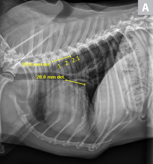
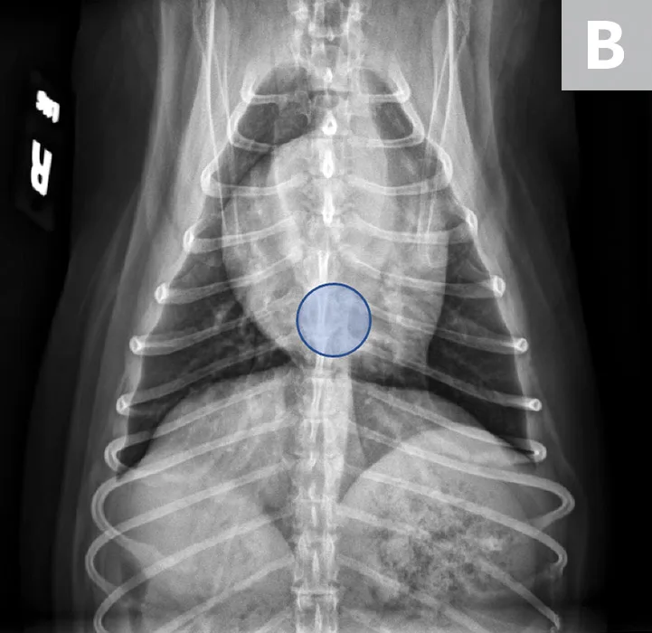
VLAS measurement in a dog with suspected stage B1 MMVD using a left lateral radiograph (A). A caliper was used to measure from the center of the most ventral aspect of the carina to the most caudal aspect of the left atrium, where it intersects with the dorsal border of the caudal vena cava. This measurement was transferred to the cranial aspect of T4 and extended caudally along the thoracic vertebrae. The number of vertebrae this line transversed was rounded to the nearest tenth to determine VLAS (2.1 VBUs). Stage B1 MMVD was confirmed via echocardiography. A redundant dorsal tracheal membrane, gastric food/foreign material, and mild T3 to T4 spondylosis deformans can also be seen. An orthogonal radiograph of the patient provides a more complete evaluation of cardiac silhouette (B); normal location of the left atrium is indicated (circle).
Stage B
Stage B includes dogs with structural heart disease that do not exhibit clinical signs of heart failure. This stage is subdivided into stage B1 (see Vertebral Left Atrial Size in a Dog with Suspected Stage B1 MMVD, above), which includes dogs with no or little cardiac remodeling that do not require treatment, and stage B2 (see Vertebral Left Atrial Size in a Dog with Suspected Stage B2 MMVD, below), which includes dogs with more advanced cardiac remodeling for which medical intervention is recommended to delay onset of congestive heart failure (CHF). Diagnostic criteria for stage B2 include a murmur intensity ≥III/VI; vertebral heart size (VHS) ≥10.5 via radiography; left ventricular internal dimension in diastole, normalized for body weight (LVIDDN) ≥1.7; and left atrial-to-aortic root short-axis ratio (LA:Ao) ≥1.6 via echocardiography.1,2 If echocardiographic measurements are not available, a general breed VHS ≥11.5 or an increase above breed-specific VHS normal values can be used to identify radiographic evidence of cardiomegaly and stage B2 MMVD.1
Case: Vertebral Left Atrial Size in a Dog With Suspected Stage B2 MMVD
A 9-year-old spayed Chinese crested was presented for evaluation of previously diagnosed stage B1 MMVD. On physical examination, a grade IV-V/VI left apical systolic murmur was auscultated. All other physical examination results were within normal limits. The patient had a history of intermittent vomiting but no other previous medical concerns.
Thoracic radiographs revealed mild, left-sided cardiomegaly (Figure 2). VHS was 11 (normal, 9.2-10.3; >10.5 is suggestive of cardiomegaly in adult dogs14). VLAS was 2.8, suggesting left atrial enlargement. Based on physical examination and radiographic findings, stage B2 MMVD was suspected. Medical therapy and continued monitoring (ie, physical examinations, thoracic radiography) were recommended to identify worsening of the condition and possible progression to CHF.
In patients with suspected stage B2 MMVD, echocardiography should ideally be performed to confirm diagnosis and determine whether pharmacologic intervention with pimobendan is warranted (based on criteria from the Evaluation of Pimobendan In dogs with Cardiomegaly study).2
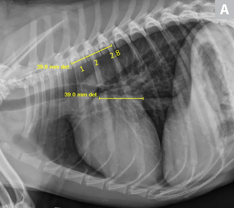
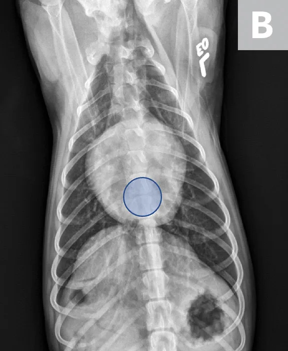
VLAS measurement in a dog with suspected stage B2 MMVD using a right lateral radiograph (A). A caliper was used to measure from the center of the most ventral aspect of the carina to the most caudal aspect of the left atrium, where it intersects with the dorsal border of the caudal vena cava. This measurement was transferred to the cranial aspect of T4 and extended caudally along the thoracic vertebrae. The number of vertebrae this line transversed was rounded to the nearest tenth to determine VLAS (2.8 VBUs). Stage B2 MMVD was confirmed via echocardiography. Other structures are unremarkable. An orthogonal radiograph of the patient provides a more complete evaluation of cardiac silhouette (B); normal location of the left atrium is indicated (circle).
Stage C
Stage C includes dogs that are or were in CHF due to MMVD but are responsive to treatment.
Stage D
Stage D includes dogs with end-stage MMVD that are in heart failure and are refractory to treatment.
Normal Vertebral Left Atrial Size Values
Although specific breed reference intervals have not been published, pilot studies suggest VLAS <2.3 correlates well with lack of echocardiographic evidence of left atrial enlargement in dogs.3,4 VLAS ≥2.3 to 2.5 suggests left atrial enlargement, and dogs with VLAS exceeding this range likely have hemodynamically important MMVD that requires treatment and thorough echocardiographic evaluation.4-6 VLAS 2.5 to 2.8 suggests stage B2 MMVD, but further evaluation via echocardiography is recommended to make a diagnosis.3 Without echocardiography, VLAS ≥2.8 to 3.1 is likely consistent with stage B2 MMVD; however, more studies are needed to determine predictive value.1,3,5,6
There has been no observed effect of body weight, sex, or age on VLAS, but there may be variation due to differences in breed vertebral conformation that are consistent with breed variations in VHS.7,8 No breed reference ranges have been established. One study found VLAS 1.8 ± 0.2 in healthy Chihuahuas; this is significantly lower than the reference range (2.07 ± 0.25) reported in a previous study.4,7 These results suggest breed-specific VLAS reference values should be determined for more accurate diagnosis.
Uses of Vertebral Left Atrial Size
VLAS provides an objective measurement of left atrial size on thoracic radiographs and is a reliable predictor of left atrial enlargement in dogs with MMVD.4,9 Although echocardiography remains the gold standard for diagnosing cardiac chamber enlargement, it may not be an option due to pet owner financial limitations, lack of patient cooperation, and/or clinician inexperience performing and interpreting echocardiography. In contrast, thoracic radiography is less expensive, more readily available, and more easily interpreted by clinicians.
Due to individual variability among dogs and lack of breed-specific reference ranges, VLAS should be used with clinical examination findings (eg, presence of murmur, typical signalment [ie, small breed, middle aged or senior]), other radiographic diagnostic findings (eg, chamber enlargement patterns), or additional imaging modalities (eg, thoracic-focused point-of-care ultrasonography, echocardiography) to diagnose MMVD.
Studies including healthy dogs and dogs with stage B1 to D MMVD found positive correlation among VLAS and LVIDDN, VHS, and LA:Ao ratio, suggesting VLAS is an indicator of cardiac remodeling and left atrial enlargement.4,5,8,10 In one study of dogs with suspected or diagnosed cardiovascular disease, VLAS was a more specific indicator of left atrial enlargement than increased VHS.3 Another study, however, found VLAS did not significantly improve detection of left atrial enlargement compared with VHS, and there was a low correlation between VLAS and echocardiographic LA:Ao ratio.11 This discrepancy may be due to differences in patient populations—only dogs with preclinical MMVD (stage B1 and B2) were investigated in the latter study.
VLAS increases based on severity of MMVD and decreases after treatment (alongside a decrease in VHS, LA:Ao ratio, and LVIDDN).10 VLAS ≥2.8 is highly specific for dogs with LA:Ao ratio ≥1.6 and therefore can help differentiate stage B1 MMVD from stage B2; VLAS 2.5 to 2.8 can help identify dogs that may benefit from echocardiography to determine whether they have reached stage B2.5,11
Noncardiac Factors That Can Affect Vertebral Left Atrial Size
Interobserver Variability
High intra- and interobserver agreement for VLAS measurements has been shown, although slight differences in measurements have been found between students and experienced clinicians.4,8,12
Abnormal Vertebrae & Intervertebral Disk Space
Abnormally shaped vertebrae (eg, hemivertebrae, fused vertebrae) are common in brachycephalic breeds and can impact VLAS measurement. Narrowed intervertebral disk spaces can also impact VLAS measurement, producing falsely increased values.
Right Versus Left Lateral Recumbency
Changes in recumbency may impact VLAS, but studies do not show significant differences.4,6 Consistent patient positioning is recommended for serial VLAS measurements, although it may not be clinically significant.
Step-By-Step: Measuring Vertebral Left Atrial Size
WHAT YOU WILL NEED
Right or left lateral thoracic radiograph
Calipers (electronic calipers can be used with digital films)
STEP 1
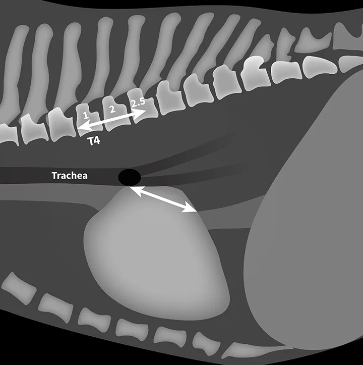
Use calipers to measure from the center of the most ventral aspect of the carina to the most caudal aspect of the left atrium, where it intersects the dorsal border of the caudal vena cava, on the right or left lateral thoracic radiograph.
STEP 2
Transfer the left atrial measurement to the cranial aspect of the fourth thoracic vertebrae (T4), extending caudally along the vertebrae at midbody position. Count the vertebrae transversed, and round to the nearest tenth (express the measurement, VLAS, in vertebral body units [VBUs]). VLAS is 2.5 in Figure 3.
Author Insight
Normal left atrial size on thoracic radiographs does not rule out cardiac disease or dysfunction. VLAS in conjunction with other diagnostics (eg, physical examination, patient history, ECG, echocardiogram) can help determine whether treatment for cardiac disease is warranted or further testing is needed based on clinical suspicion of cardiac involvement in disease.